


Shocking the cellular world: Engineers’ collaborative work discovers force signature of cells undergoing electroporation
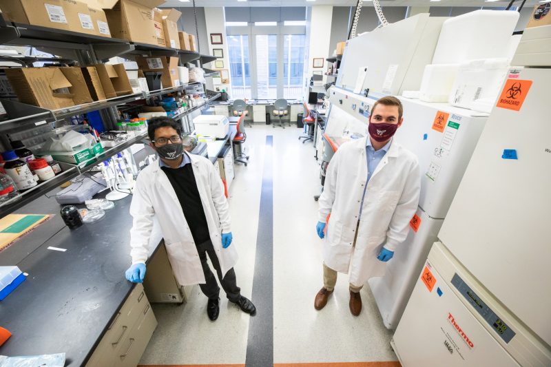
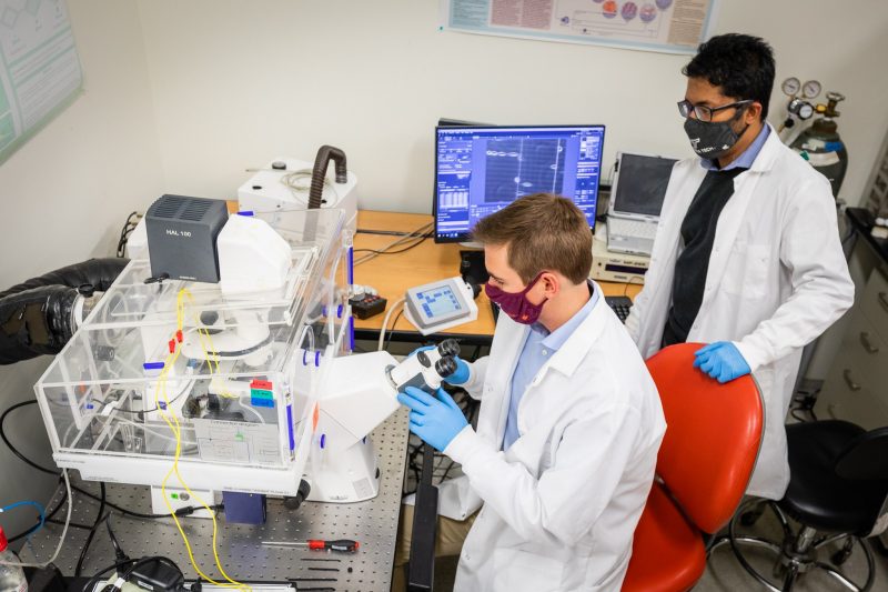
Aniket Jana (left) and Philip Graybill (right) conduct research in engineering labs.
The idea to collaborate came to Rafael Davalos and Amrinder Nain in a conversation on the stairs. Davalos, the L. Preston Wade professor in biomedical engineering and mechanics, and Nain, associate professor of mechanical engineering, both have research space at the Kelly Hall location of the Institute for Critical Technology and Applied Science at Virginia Tech.
"We crossed paths on the second-floor staircase landing," said Nain. "I said, 'I do force measurement of single cells, you use electric fields on cells.’ We knew of each other's research and wondered what we could do with our combined expertise.'”
After brainstorming, they came up with a plan to see if they could study the forces exerted by a cell undergoing electroporation.
Combining their respective expertise in cell force measurement and electroporation in a single study published as the cover feature of American Chemical Society’s (ACS) flagship ACS Nano journal, Nain and Davolos discovered a mechanical response from cells that may open doors to medical advances. They discovered an unusual biphasic response that could change the way the medical field understands and approaches techniques using electroporation.

Electroporation has been used in many medical applications, such as gene transfection and electrochemotherapy, since the 1980s. The basic aim is to apply a voltage to a cell; that voltage then pulses across the cell’s membrane and at certain magnitudes causes pores, or “holes,” to form in it. In this study, the researchers improved upon an established method of electroporation, in which medicines or genes are injected into holes formed in a cell’s membrane.
While much is known about the methods of creating these openings, the cell’s mechanism for resealing has remained largely unexplored. According to the researchers, by investigating the mechanical responses as cells contract following electroporation, they could gain a deeper understanding of how cells adapt their structure to promote their recovery. Unlocking cell recovery could in turn advance knowledge of cell mechanics and membrane permeability, as well as improve techniques harnessing electroporation, like gene transfection, electrochemotherapy, and other cancer treatments.
In the study, Davalos and his graduate students in the Bioelectromechanical Systems Laboratory, engaged in in vitro electroporation, a method of administering electrical pulses to tissue to treat cancer. Electroporation was applied to numerous types of cells, including human glioblastoma and mesothelioma cells. Nain and his graduate students in the Spinneret based Tunable Engineered Parameters (STEP) Laboratory engaged in creating precisely controlled, suspended fibrous environments. This brought a unique perspective to electroporation, enabling careful observation of cellular mechanical response throughout the process.
In their experiments, the researchers were able to culture cells on suspended nanofibers, rather than on the flat bottom of a petri dish, which allowed them to mimic the native fibrous environment found inside the body. In Nain’s lab, researchers used nanonet force microscopy, a method pioneered by Nain, to measure cell forces. Cells attached to the nanonets and bent the flexible fibers, enabling the measurement of their contractile forces. In Davalos’ lab, the nanonets were assembled in a custom microfluidic device housing two electrodes.
“Cell properties and forces can be hard to understand intuitively,” said Philip Graybill, a mechanical engineering doctoral student and first co-author on the study publication. “How much force is a nanonewton anyway? Other methods of characterization tend to be somewhat abstract, but in this study, the nanofibers provided a visually fascinating method to investigate cell behavior. Images of the nanofibers bending under the force of a cell brought these values to life.”
“Cell forces have been studied before, to understand various biological phenomena, but never in the context of electroporation,” added Aniket Jana, a mechanical engineering doctoral student and first co-author on the study. “Our approach to monitor cell force dynamics provides a direct way of understanding the physical recovery mechanisms of cells following electric field treatments.”
The unique experimental setup led the team to their fundamental discovery. When high electric fields were applied, cellular forces decreased, which was concurrent with the formation of pores in the cell membrane. Cell force recovery coincided with membrane resealing, but the recovery showed an unusual biphasic response: increase and decrease in forces before the cells finally recovered their original contractility. Applying forces (contractility) is fundamental to the nature of cell. Cells exert this force, mediated by the cytoskeleton, to divide themselves, migrate, or heal wounds.
To the researchers, this biphasic response made it clear that cytoskeletal dynamics play a significant role in cell shape and force recovery.
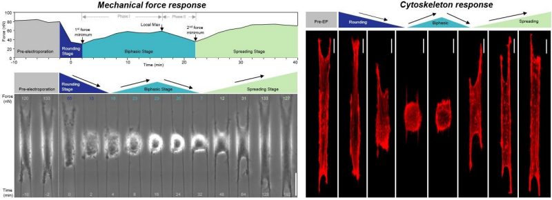
Nain explained that cell membrane disruption is linked to the loss of contractility-bearing cytoskeleton inside the cell. He described the cytoskeleton as a dynamic and responsive meshwork; its re-establishment is required for recovery of the cell’s contractility and shape.
“It is remarkable how the cell cytoskeleton can fully recover within just a couple of hours, despite extensive damage caused by electric fields,” Jana said. “This shows the strong will of cells to live even after extreme perturbations. Such incredible cell adaptability in our suspended nanofibers further highlights how cancer cells can evade electric field treatments and still continue on metastasizing.”
“It was amazing to see just how dynamic and responsive cells really are after electrical disruption,” added Graybill. “It was exciting to find that the loss of contractile force measured in one cell type also occurred in other cell types, because this suggested that this behavior is consistent across many cell types.
"This was exciting, because a better understanding of cell recovery may improve techniques where cell recovery is desirable, such as in gene transfection, electrofusion, electrochemotherapy, or where cell recovery is undesirable, such as in cancer treatments, where it’s preferable for the cancerous cells to die.”
The researchers believe their new understanding of cell contraction and recovery has important implications for electroporation’s use in various applications, including molecular medicine, genetic engineering, and cellular biophysics. Nain said that the biphasic response they observed could enable the injection of larger particles into the cell, without having to use a higher electric voltage.
The cell membrane is like an insulator, and when you apply a voltage, the electrical pulse travels across the membrane, explained Davalos. At a critical voltage, pores form on the cell. The magnitude of the electric field controls whether or not pores will form in the membrane, while the various pulse parameters dictate the size of the particle that can be put into the cell.
Trying to increase those parameters could affect the particle size one can inject, but the higher the electrical voltage, the more likely the cell is to die. There is also a very narrow window where pores are open and able to receive materials before the cell reseals. The overall goal of electroporation is to disrupt the cell so that substances – such as medicine or DNA – can be injected into it. Identifying and understanding how long the disruption lasts is thus very important.
“This shows the strength of collaboration across disciplines,” Nain said. “I knew about the electric field research by Davalos and wondered how we could integrate nanonet force microscopy, the cell force measurement platform developed in my lab, with electrical fields. Together, we have discovered a new phenomenon in a well-established field.”
In Nain and Davalos’ study, the cells attached to suspended fibers were able to sustain high electrical voltages while enabling the precise measurement of forces. This holds great promise to deliver macromolecules in the cells efficiently, they said.
“This platform has broadened that reversible window,” Davalos said. “This is exciting because the opportunity for introducing larger particles increases without the risk of killing the cell. This could have major implications in genetic engineering and molecular medicine. Achieving this in realistic fibrous environments opens new possibilities for translating the technology.”
The research was conducted by an interdisciplinary team from multiple engineering backgrounds. Philip Graybill and Aniket Jana, Ph.D. students in mechanical engineering, are equal authors on the publication, and are joined by Rakesh Kapania, Norris and Laura Mitchell Professor in the Kevin T. Crofton Department of Aerospace & Ocean Engineering.
-Written by Laura McWhinney and Alex Parrish